
Acad J Health. 2024; 2(1): 20-23 | DOI: 10.14744/ajh.2024.32932
Wilsons Disease: Brain Magnetic Resonance Imaging
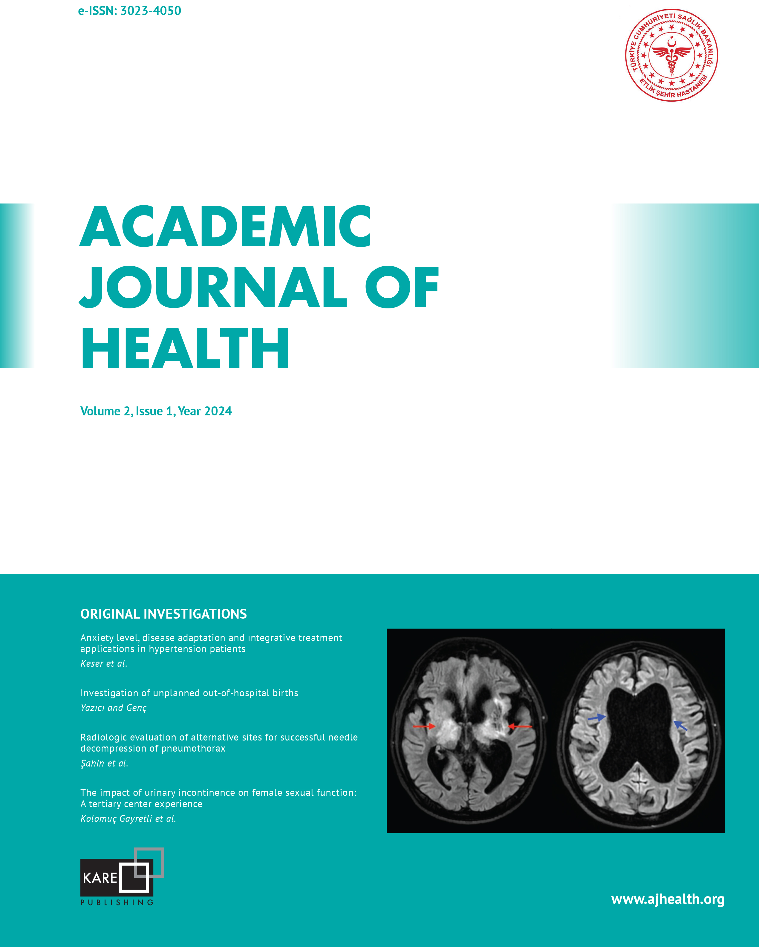
Emine Ayten Aksoy Keleţ, Sümeyya Duran Kaymak, Berna Turhan, Rasime Pelin KavakDepartment of Radiology, Ankara Etlik City Hospital, Ankara, TürkiyeWilson’s disease (WD) is an autosomal recessive systemic disorder that causes copper accumulation and toxicity in multiple organs. Various neurologic symptoms, such as rigidity, tremor, bradykinesia, dystonia, chorea, dysarthria, and dysphagia, are observed in the central nervous system involvement of WD. Magnetic Resonance Imaging (MRI) usually shows increased T2-weighted signals in the basal ganglia, mesencephalon, and pons. MRI is a valuable, non-invasive imaging technique for the diagnosis and follow-up of central nervous system involvement in life-threatening WD. In this case report, we will present cranial MRI findings of WD with cranial involvement.
Keywords: Hepatolenticular degeneration, Wilson's disease, magnetic resonance imagingCorresponding Author: Emine Ayten Aksoy Keleţ, Türkiye
Manuscript Language: English
Manuscript Language: English